HEALTH 2019: Merit Health introduces new mammography technology
Published 11:34 pm Saturday, April 27, 2019
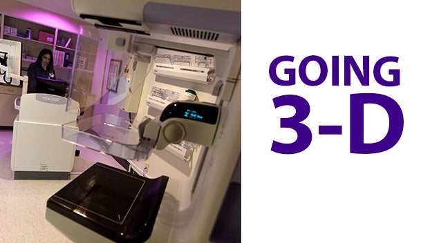
- Merit Health Natchez mammography technologist Sharree Griffin readies for the next patient to get a mammography using the hospital’s new technology. (Ben Hillyer / The Natchez Democrat)
NATCHEZ — A lot has changed in the world of mammography in the 29 years that Sharree Griffin has provided the diagnostic tool for patients in the region.
A mammography technologist for the Merit Health Natchez, Griffin first performed mammograms using technology developed by Xerox.
“I started on a Xerox when the patient laid on her side,” Griffin said. “Then we went to film screen and then we went to digital.”
On April 24, the next advancement in mammography technology — three-dimensional mammography — was introduced to patients in the Miss-Lou at Merit Health Natchez.
“It has come a long way since those early days,” Griffin said.
Not only do patients no longer have to lie on a cold table as they did in the 1970s, but they can also now get a more definitive diagnosis with less discomfort, said Susan Grayson, Merit Health director of medical imaging.
“We are extremely pleased to add this technology, the only one of its kind in the Miss-Lou,” CEO Lance Boyd said.
The new diagnostic tool, Boyd said, provides better and earlier breast cancer detection for patients than the older two-dimensional technology.
Unlike traditional 2-D mammograms, the new technology takes several images of the breast to create a three-dimensional image.
“Instead of four pictures you are getting approximately 400 pictures taken during the exam in the same amount of time,” Grayson said.
The 3-D scan provides a more detailed look with approximately the same radiation dose, Grayson said.
Officials from the American College of Radiology said 3-D mammography exams are shown to be an advancement over 2-D exams, with higher cancer detection rates and fewer patient recalls for additional testing.
“The technology also enhances the clarity of the images … and will result in better piece of mind for our patients,” Grayson said.
During a 3-D exam, an X-ray arm sweeps in a slight arc over the breast, taking multiple images. A computer then converts the image into a stack of thin layers, allowing the radiologist to view the breast tissue one layer at a time.
Grayson said patients might not notice much difference between the 3-D technology and the conventional 2-D technology, in that the exam will take about the same amount of time and the same positioning. The most significant difference is in the information available to the radiologists reading the exams.
Radiologist Dr. Norwood Smith, who works for Jackson Radiology Associates and provides services to Merit Health Natchez agrees.
“This technology is a game-changer in the fight against breast cancer,” Smith said. “I commend the hospital for investing in it for the community.”
More News


SportsPlus
Where to Watch Nashville Predators vs. Winnipeg Jets on TV or Streaming Live – November 23
NHL action on Saturday includes the Nashville Predators (6-11-3) hosting the Winnipeg Jets (17-3) at Bridgestone Arena, starting…
Predators vs. Jets Injury Report Today – November 23
Entering a game against the Winnipeg Jets (17-3), the Nashville Predators (6-11-3) will be monitoring two players on…
How to Watch Top 25 College Basketball Games – Saturday, November 23
There are four games on the college basketball schedule on Saturday that feature a ranked squad. That includes…
Best Bets for NCAA Basketball Picks Against the Spread for Saturday, November 23
The college basketball slate on Saturday, which includes the Saint Francis (PA) Red Flash taking on the Georgetown…
How to Watch the NBA Today, November 23
On a Saturday NBA card that includes seven exciting contests, the Golden State Warriors versus the San Antonio…
-
Polls
 Loading ...
-
-
Special Sections
